Imaging
View in the inner world
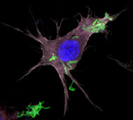
Imaging plays a crucial role to understand inflammatory processes in cells and tissue cultures. Within common approaches fluorescent and bioluminescent samples give an insight into three dimensional space, structure and temporal dynamic. Particularly high-resolution microscopy reveal molecular mechanisms and alterations which determine acute inflammation.
Further non-invasive radiological and nuclear medicine imaging offer new insights into living organisms. In this project the Optical Imaging Center Erlangen (OICE) – a cooperation of FAU and Max-Planck-Institute for the science of light – and the Preclinical Imaging Platform Erlangen (PIPE) offer customized to the CRC1181 scientific projects progressive imaging infrastructure and solutions.
This central project opens up new ways to investigate inflammatory diseases and consequent therapies, allowing the FAU to establish itself as a worldwide leader in inflammatory research